Article Text

Abstract
Objectives To assess the intravascular volume status using bedside ultrasonography of the internal jugular vein in the ED & to compare the results of two different methods, one qualitative and the other quantitative.
Background Estimation of intravascular volume status is an important parameter both for initial assessment and to evaluate the response to treatment in critically ill undifferentiated patients especially in an ED setting.
Point of care ultrasound has been gaining popularity in the practice of EM. Ultrasound of the inferior vena cava (IVC) is used in estimating the Intra vascular volume, and is part of the RCEM's level 2 ultrasound training. IVC can be difficult to visualise in some patients. Internal Jugular Vein (IJV) is more easily accessible. Ultrasonography of the IJV has been described in the literature both as a qualitative and a quantitative tool to predict intravascular volume status.
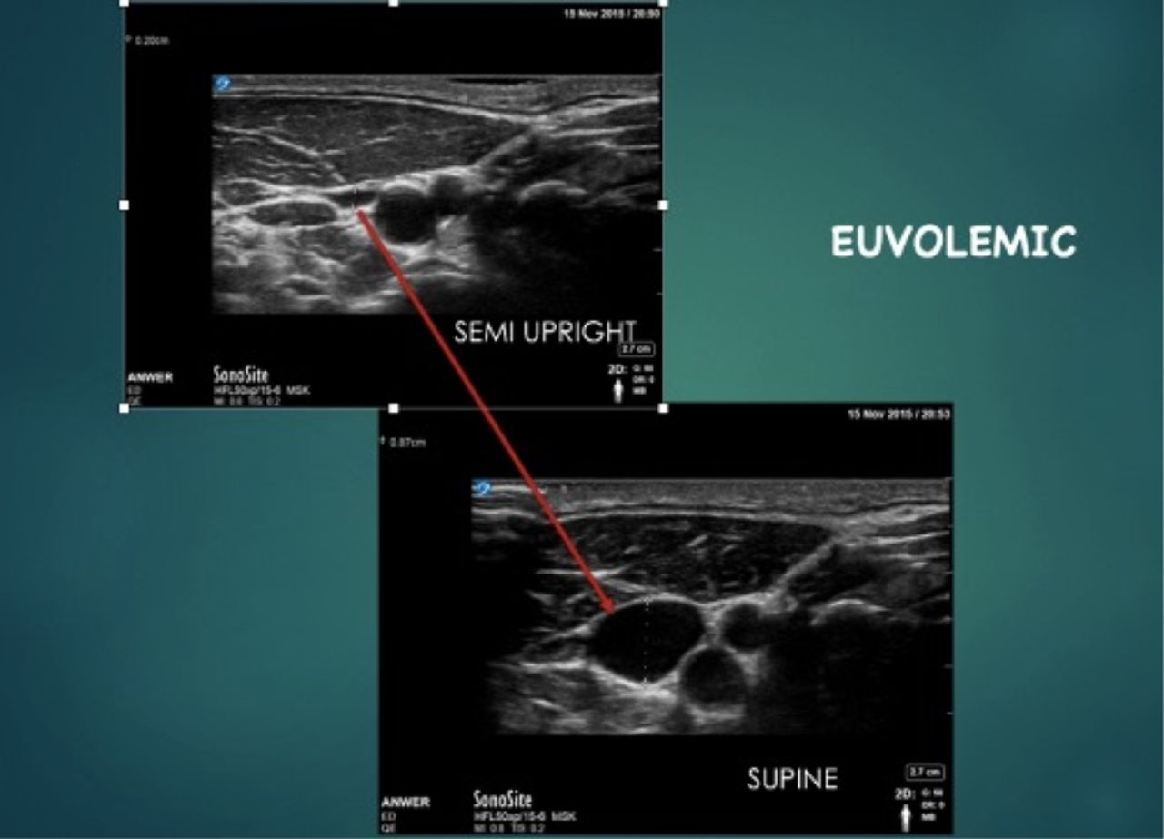
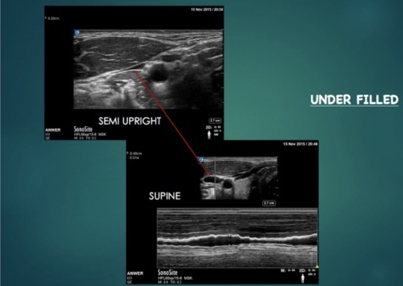
Methods A prospective feasibility study at the ED of a tertiary hospital. We used a convenience sample. In phase-1 we scanned 15 healthy volunteers and in phase-2 we scanned 21 acutely unwell adult patients from Oct to Nov 2015. The appearance of the IJV was noted in first 45 degrees and than in supine position as described by Lipton, 2008. Than the aspect ratio (height/width) of the IJV was measured in supine position as described by Keller et al, 2009. We used a high frequency linear probe at a depth of 3.5–4 cm. The EP performing the ultrasound was blinded to the clinical information. The findings of the ultrasound were kept blinded from the attending EP. Results of the ultrasound were retrospectively compared with the final clinical judgment of the attending EP using clinical data to assess the fluid status of the patient as recommended by NICE Guidelines on intrvenous fluid therapy, 2013.
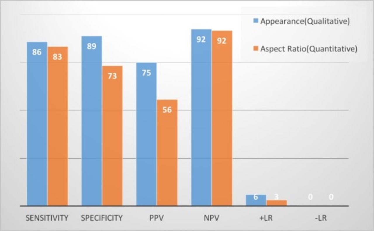
Results The results showed that for assessing the intravascular volume status bedside ultrasonography of the IJV byaspect ratio technique has a Sensitivity of 83.33%.
▸ Specificity of 73.33
▸ PPV of 55.56
▸ NPV of 91.67
▸ LR+of 3.12
▸ LR– of 0.23
▸ Whereas by appearance technique
▸ sensitivity of 85.71%
▸ Specificity of 88.71
▸ PPV of 75.0
▸ NPV of 92.31
▸ LR+of 6
▸ LR– of 0.17

- Trauma