Article Text

Abstract
Background The optimal management of minor head injured patients with brain injury identified by CT imaging is unclear. Some guidelines recommend routine hospital admission of GCS13–15 patients with traumatic brain (TBI) injury identified by CT imaging. Others argue that selected lower-risk patients can be discharged from the Emergency Department (ED).
Objective To estimate the risk of death, neurosurgery and clinical deterioration minor head injured patients with TBI identified by CT imaging, and assess which factors affect the risk of these outcomes.
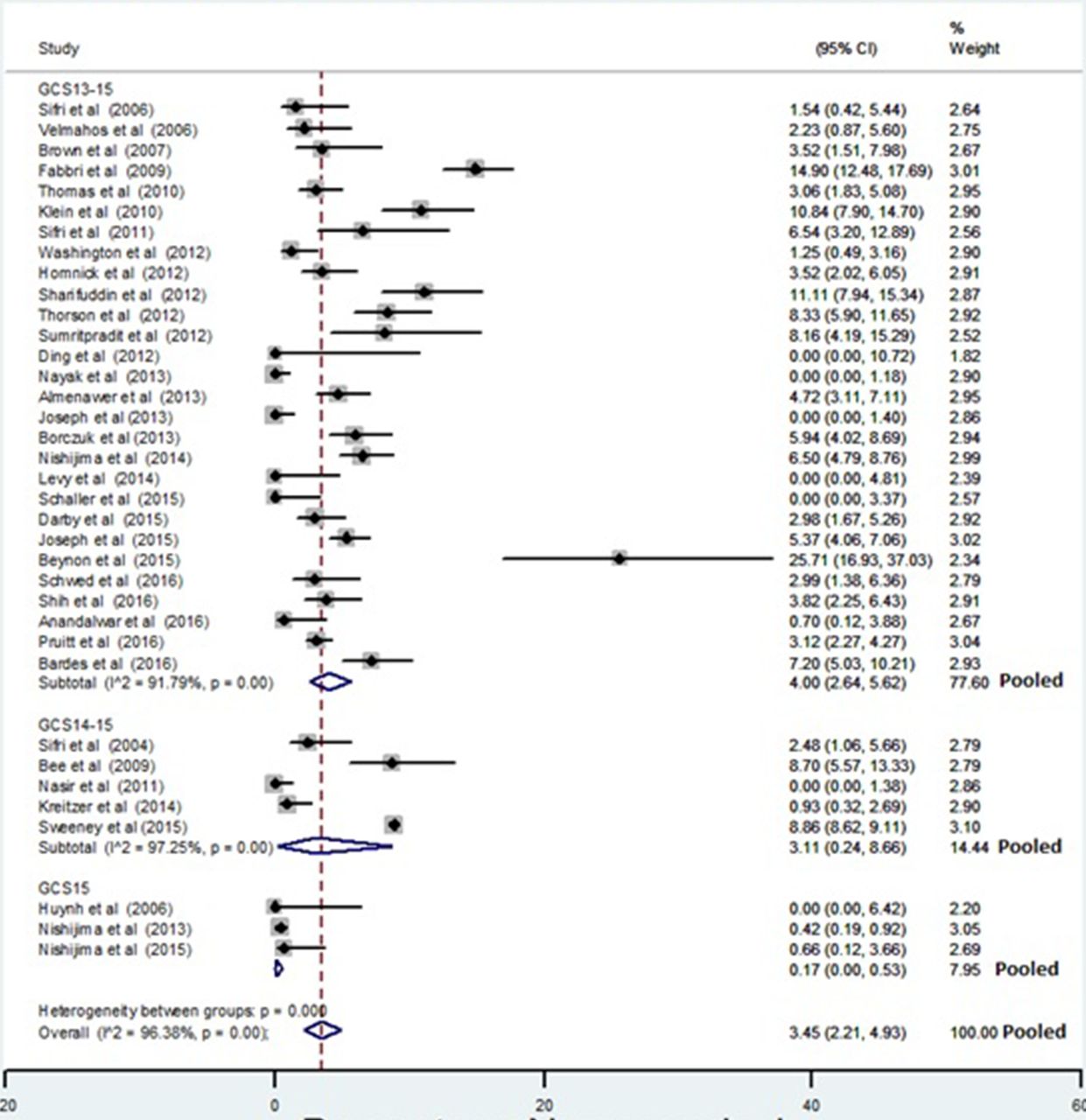
Risk of neurosurgery stratified by the initial GCS of the study population
Methods A systematic review and meta-analysis adhering to PRISMA standards of reporting. Four electronic data bases and a range of additional literature were searched using a highly sensitive search strategy. Study selection was performed by 2 independent reviewers. Meta-analysis using a random effects model was undertaken to estimate pooled risks of: clinical deterioration, neurosurgery and death. Meta-regression was used to explore between study variation in outcome estimates using study characteristics. Factors assessed by individual studies as affecting the outcomes of interest were recorded and pooled within study risk factor effects were estimated where possible.
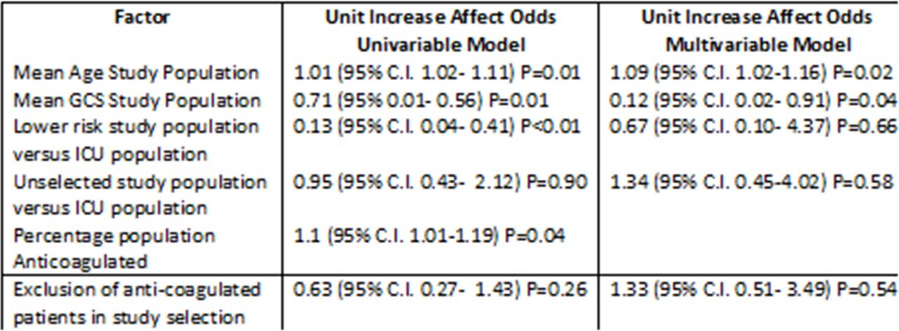
Results 4431 studies were identified by the search strategy, of which 123 studies were fully retrieved and 49 primary studies and 5 reviews met the inclusion criteria. The estimated pooled risk of the outcomes of interest were: clinical deterioration 11.7% (95% CI:11.7 to 15.8; neurosurgery 3.5% (95% CI:2.2% to 4.9%); death 1.4% (95% CI:0.8% to 2.2%). A large degree of between study variation in the estimates of the outcomes was identified. Multivariable meta-regression of study characteristics identified that mean age of the study population and mean initial GCS accounted for up to half of the variation in reported study outcomes. Within studies the following factors were found to affect the risk for these adverse outcomes: age; severity of injury; type of injury; initial GCS; anti-coagulation; anti-platelet medication; and injury severity scoring. When univariable within study risk factor effect estimates were pooled patients with isolated subarachnoid haemorrhage had an odds ratio of 0.19 for deterioration compared to other injury types.
Meta-regression of study factors predictive of neurosurgery
Conclusion Minor head injured patients with brain injury identified by CT imaging have a clinically important risk of serious adverse outcomes. Research has identified the possible factors that affect this risk. However, these factors need to be incorporated into a validated multivariable prognostic model before low-risk patients can be reliably identified clinically and triaged to lower levels of care.
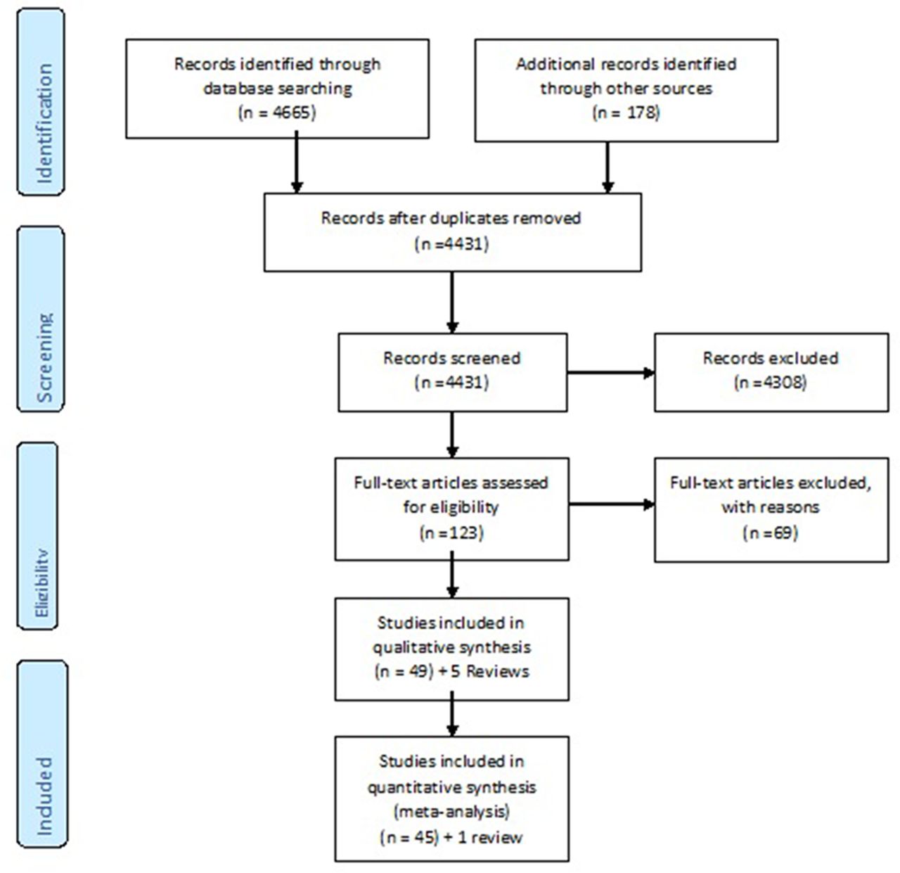
PRISMA flow-diagram showing selection of studies for inclusion in the systematic review