Article Text

Abstract
Background: Rapid sequence induction (RSI) is increasingly used by emergency physicians in the emergency department. A feared complication of the technique is the inability to intubate and subsequently ventilate the patient. Current drills based on anaesthetic practice may be unsuitable for use in the emergency department.
Objective: To construct a drill for failed adult intubation in the emergency department.
Methods: Literature review and consensus knowledge.
Results: A drill for failed adult intubation in the emergency department is given.
Summary: Failure to intubate following RSI in the emergency department is a feared complication. Practitioners must have a predetermined course of action to cope with this event. The guidelines presented here are tailored for use by the emergency physician.
- rapid sequence induction
- failed intubation
- laryngoscopy
- RSI, rapid sequence induction
- LMA, laryngeal mask airway
Statistics from Altmetric.com
Securing the airway by tracheal intubation is an essential component of resuscitation in the emergency department.1 Though some patients can be intubated without the use of drugs, many patients requiring intubation will still have some degree of airway reflexes and will require pharmacological adjuncts to overcome these to facilitate tracheal intubation. The most commonly used technique to achieve this is rapid sequence induction (RSI),2 which consists of: a period of preoxygenation, the administration of a short acting intravenous hypnotic (often an anaesthetic induction agent) followed by a neuromuscular blocking drug, for example suxamethonium. During this sequence, as consciousness is lost, cricoid pressure is applied to reduce the risk of regurgitation and aspiration.
In North America, the alternative term “rapid sequence intubation” is used when the same process is used in the emergency department. This is to reinforce the concept that tracheal intubation is the primary aim for emergency department patients, whereas for anaesthetic patients RSI is an initial part of the anaesthetic process.
RSI is an important skill, which exists within the domain of emergency medicine practice (Graham CA et al, annual scientific meeting of the Faculty of A+E medicine, London, December 1999, Mackay CA et al, annual scientific meeting of the Faculty of A+E medicine. London, December 1999 and references 1,3,4). Therefore all clinicians involved in the use of RSI must be adequately prepared to deal with the possibility of a failed intubation.5 This paper proposes a guideline such a scenario occurring in adult patients in the challenging environment of the emergency department.
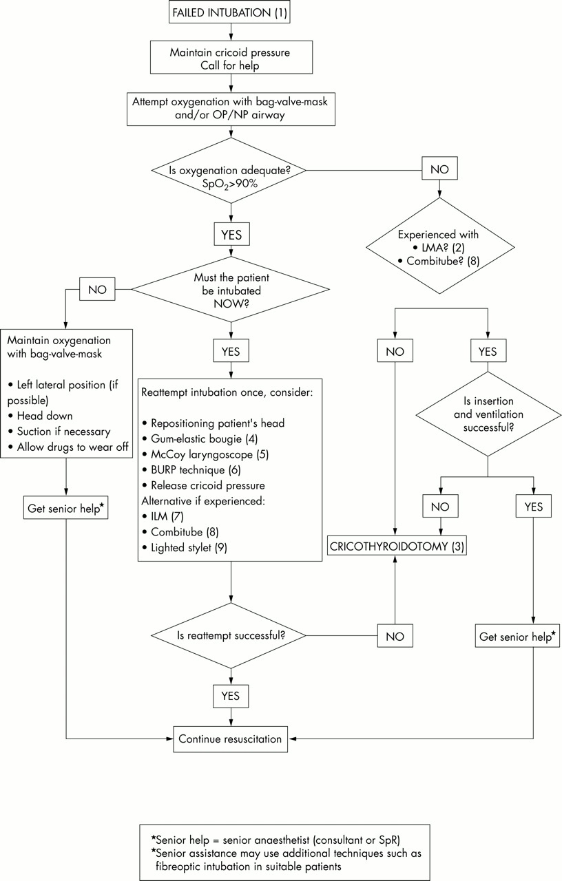
FAILED INTUBATION DRILL IN THE EMERGENCY DEPARTMENT
Guideline notes
-
The case mix of patients in the emergency department is difficult and clinically challenging. Personal and equipment preparation are essential components in the management of a failed intubation. Essential equipment should be stored in the resuscitation area and be clearly identifiable as difficult airway equipment (box 1).
-
The laryngeal mask airway (LMA) may be a lifesaving adjunct during a failed intubation. Successful insertion is not affected by the presence of factors used to predict a difficult intubation.6–8 Correct placement of an LMA is compromised by the application of cricoid pressure.9–11 Consequently release of cricoid pressure will probably be necessary, and the risk of subsequent regurgitation and aspiration must be balanced against achieving ventilation in these circumstances. Even a correctly situated LMA does not guarantee against aspiration, but the risk is small.12 Furthermore, it allows better ventilation than with a facemask and self inflating bag.13 Cricoid pressure can be reapplied once the LMA is in situ. Although this does not affect the position of the LMA 14 it will reduce the efficacy of ventilation.15
Box 1 Minimum essential equipment (in addition to standard airway equipment)
-
Microlaryngoscopy endotracheal tubes, sizes 5 mm and 6 mm
-
Tracheostomy tubes (cuffed), sizes 5–8 mm
-
McCoy laryngoscope
-
Gum elastic bougie
-
Needle cricothyroidotomy set (for jet ventilation)
-
Cricothyroidotomy set (for placement of tracheostomy tube)
-
LMAs sizes 3–5 OR Combitube
Desirable equipment
-
Intubating laryngeal mask airway
-
Lighted stylet
-
Fibreoptic bronchoscope
-
-
Cricothyroidotomy. All emergency departments where RSI is performed should have a cricothyroidotomy set immediately available. The RSI practitioner should know how to use it!
-
The use of a bougie is recommended when the view of the larynx is poor.16–18
-
The McCoy laryngoscope reduces the incidence of poor laryngoscopic views to less than 5% in patients with potential cervical spine injuries.19
-
The BURP technique is when cricoid pressure is applied Backwards, Upwards, to the Right and with Pressure.20 It has been suggested that this may improve the view at laryngoscopy. As most assistants applying cricoid pressure stand on the right of the patient they have a tendency to push to the left instead.
-
The intubating LMA has been used to facilitate intubation in patients where direct laryngoscopy has failed in the emergency setting.21 However, it requires considerable training and ongoing practice and few emergency physicians will be familiar with its use. It is mentioned here for those physicians already competent in its usage.
-
The Combitube is a double lumen tube that combines the functions of a tracheal tube and an oesophageal obturator airway. It is inserted blindly, and can be used with the neck in neutral alignment.22 An alternative method of securing the patients airway or blindly intubating the patient may reduce the number of cricothyroidotomies (Morton T, annual scientific meeting of the Faculty of A&E medicine, London, December 1999). The Combitube has the advantage of itself securing the airway. It has therefore been recommended as an adjunct for use in the cannot intubate/ventilate scenario.23,24
-
The lighted stylet is a relatively new addition to emergency care in North America but is not yet in general use in the UK.
Action card for failed intubation in the emergency department.
DISCUSSION
RSI in the emergency department presents a number of unique problems as the patients; indications and degree of urgency are very different to those encountered by anaesthetists in the operating department. As a consequence the incidence of failed intubation is much higher. For example, the incidence of failed intubation among US anaesthetists in the controlled environment of the theatre suite is 0.05%–0.35%.25 In contrast in the emergency setting the incidence has been reported to be 1%–2%26 with repeated attempts at laryngoscopy being required in 5%–20% of cases.5,27 As a result the incidence of cricothyroidotomy in US emergency departments is 0.5%–1.2%, 28–31 far higher than in the environment of the operating room. There are no comparative data from the UK, but we suspect that the incidence is very much lower. This is probably because of greater availability at present, of experienced anaesthetists or emergency physicians with anaesthetic training.
Patient factors
Certain groups of patients can be predicted as having an increased likelihood of being difficult to intubate. This may be as a result of their presenting condition; for example facial trauma or a pre-existing morbidity condition such as ankylosing spondylitis (box 2). Anaesthetists have described a number of clinical assessments to try and predict difficulty with intubation. However, the majority of these require both time and a cooperative patient, neither of which may be present in the victim in the emergency department.32,33 One situation guaranteed to make intubation difficult is the presence of a semi-rigid collar, head blocks and tape to immobilise the cervical spine. In nearly two thirds of these patients, the larynx will not be visible on laryngoscopy. The use of manual in line stabilisation will reduce the incidence of poor view to around 20% as a result of improved mouth opening.18,31
Box 2 Conditions likely to predispose to a difficult intubation
Obvious syndromes/conditions
-
Pierre-Robin syndome
-
Acromegaly
-
Pregnancy
-
Ankylosing spondylitis
-
Rheumatoid arthritis
Obvious anatomical conditions
-
Obesity
-
Bull neck
-
Prominent teeth
-
Poor dentition
Obvious trauma
-
Maxillary facial trauma
-
Neck trauma
-
Laryngeal trauma
-
Airway obstruction
In addition the types of patients requiring airway intervention in the emergency department are likely to present problems (box 3).
Box 3 Airway/ventilation problems associated with serious illness and injury
-
Pre-oxygenation may be impossible or ineffective
-
Positioning for intubation may be difficult if the cervical spine is immobilised32
-
The airway may be partially obstructed by trauma, blood, vomitus or secretions
-
The patient may be uncooperative
-
They patient may already be hypoxic or haemodynamically compromised
-
It may be impossible to predict whether the patient is likely to represent a difficult intubation
Despite these problems airway control may be urgently required. Consequently, such patients are at high risk of suffering a complication of an attempted intubation 34 (box 4).
Box 4 Complications of attempted intubation in the emergency department
-
Failure to intubate
-
Hypoxia
-
Unrecognised oesophageal intubation
-
Aspiration of stomach contents
-
Hypotension
-
Awareness
-
Arrhythmias
-
Cardiac arrest
Clinical indications for RSI
The patients in whom RSI is likely to be used can be divided into two main groups. An example of the first would be a patient who has taken an overdose, is now comatose, cardiovascularly stable and maintaining a patent airway. Protection of the airway is desirable but not required immediately. If subsequent attempts at tracheal intubation fail, the effects of hypnotics and neuromuscular blocking drugs can be allowed to wear off, with the patient tipped head down and in the left lateral position, while cricoid pressure is maintained. Many current anaesthetic guidelines are applicable in these circumstances.24
The second group of patients are those in whom it is essential to secure the airway as part of the resuscitation process. This is usually as a result of a compromised airway or ventilatory failure (box 5).
Box 5 Examples of patients requiring emergency RSI
-
Isolated head injury. Hypoxic, GCS 5, facial injury, blood in the pharynx, masseter spasm
-
Chest injury requiring urgent ventilation (for example, bilateral flail segments; pulmonary contusion; drained haemopneumothoraces with hypoxia despite adequate drainage and supplemental oxygen)
-
Asthma. Exhausted asthmatic on maximal therapy
-
Status epilepticus unresponsive to other therapy
Once an attempt at intubation has commenced, the ideal final outcome is a cuffed tube in the patient's trachea. Clearly the option of allowing the effects of drugs administered to wear off is neither practical nor desirable in these patients.
Equipment
Emergency departments are generally less well equipped to deal with tracheal intubation than anaesthetic rooms, particularly when there are predicted or unexpected difficulties. Anaesthetic rooms will contain various laryngoscopes, bougies and alternative airway devices, along with immediate access to a “difficult intubation box” (see box 1) or its equivalent. This is not the case in many emergency departments in both the UK (Morton T, annual scientific meeting of the Faculty of A&E medicine, London, December 1999) and US 35 and is of serious concern considering the greater potential for encountering a difficult intubation in this setting.
Furthermore, it is common practice to rely on intravenous agents to facilitate intubation in the emergency department, as anaesthesia machines are not always immediately available. This precludes the use of inhalational agents to render patients unconscious and abolish laryngeal reflexes, which may be of value where intubation can be predicted to be difficult and there is the need to preserve spontaneous ventilation.
Skills
Few would argue that the ideal emergency airway practitioner is someone highly skilled in airway techniques, emergency resuscitation and regularly practised. In the UK such skills are often split between anaesthetic and A&E doctors. In addition, in most hospitals on call anaesthetists are expected to help in the resuscitation room, but other commitments in theatre, or ICU means that they may not be able to provide experienced help at short notice. Even in those circumstances where help is available it is unlikely to be quick enough for truly emergent situations such as the “can't ventilate, can't intubate” scenario. Therefore it is absolutely essential that emergency physicians develop strategies for managing the emergency airway. This should be addressed during specialist registrar training, but is often not. Additional training on courses such as the National Airway Emergency Course (Morton T, annual scientific meeting of the Faculty of A&E medicine, London, December 1999) with focused secondments in anaesthesia are also required.
Current guidance for failed intubation
The use of RSI is common in anaesthetic practice where there may be doubts as to the presence or absence of stomach contents (for example, a patient with an acute abdomen, or one who has recently eaten). This occurs in many elective conditions (for example, gastric outflow obstruction, obesity, pregnancy or oesophageal reflux), and is assumed in nearly all emergency patients.
It is not then surprising that “drills” have been developed for management in the event of failed intubation in anaesthetic practice. Indeed a failed intubation drill is given in virtually all anaesthetic texts. However, the protocols vary widely with some texts containing several different drills to cope with different circumstances.36 Drills advocate waking the patient up, 37,38 place patients in the left lateral position 36 or do not result in a secured airway.39 Consequently, many are unsuitable for use in the emergency department.
ATLS teaching40 advocates the use of pharmacological adjuncts for intubation but the airway algorithm fails to outline alternative methods of achieving a successful intubation, and does not indicate clearly when a surgical airway is needed. Unfortunately, some emergency texts on airway management fail to describe a failed intubation procedure.41 These flaws make the appropriateness of available protocols questionable in the emergency department setting.
Walls' “Airway Course”26 contains a failed intubation drill for the emergency setting. In the airway course algorithm it is implicit that once the decision to intubate has been made, the operator will continue with the process until the airway is secured. Our guideline differs in that it identifies those patients in whom it is desirable to allow anaesthesia to wear off in order to arrange a further planned attempt at intubation (that is, those patients in whom intubation is not immediately required and who are easy to oxygenate).
In the UK, it is not uncommon for relatively junior members of the anaesthetic team to attend the emergency department during a resuscitation. For non-RSI practitioners leading the resuscitation team the guidelines presented here may assist them and their anaesthetic colleagues in case of a failed intubation. However, the drill is most useful for the emergency physician skilled in RSI who deals with the challenging group of patients presenting to the emergency department.
SUMMARY
RSI is an important skill for emergency physicians. The patient population is different from that seen in routine anaesthetic practice and its management may therefore differ. We propose these guidelines to specifically address failed intubation in the difficult group of patients presenting to the emergency physician.
Acknowledgments
We wish to thank Dr J Nolan for his help in preparing this paper.
Funding: none.
Conflicts of interest: none.
REFERENCES
Linked Articles
- Primary Survey