Article Text

Abstract
Background To improve the ease and safety of cricothyroidotomy especially in the hand of the inexperienced, new instruments have been developed. In this study, we compared a new indicator-guided puncture technique (PCK) with standard surgical technique (ST) regarding success rate, performance time and complications.
Methods Cricothyroidotomy in 30 human cadavers performed by 30 first year anaesthesia residents. The set chosen for use was randomised: PCK-technique (n=15) and ST (n=15). Success rates, insertion times and complications were compared. Traumatic lesions were anatomically confirmed after dissection.
Results The ST-group had a higher success rate (100% vs 67%; p=0.04). There was no difference in time taken to complete the procedure (PCK 82 s. vs ST 95 s.; p=0.89). There was a higher complication rate in the PCK-group (67% vs 13%; p=0.04). Most frequent complication in the PCK-group was injury to the posterior tracheal wall (n=8), penetration to the oesophageal lumen (n=4) and injury to the thyroid and/or cricoid cartilage (n=5). In the ST-group in only 2 cases minor complications were observed (small vessel injury).
Conclusions In this human cadaver study the PCK technique produced more major complications and more failures than the ST. In the hand of the inexperienced operator the standard surgical approach seems to be a safe procedure, which can successfully be performed within an adequate time. The PCK technique cannot be recommended for inexperienced operators.
- Cricothyroidotomy
- difficult airway management
- puncture technique
- standard surgical technique
- surgical airway
- airway
- anaesthesia
- management
- training
- equipment evaluation
- emergency departments
This is an Open Access article distributed in accordance with the Creative Commons Attribution Non Commercial (CC BY-NC 3.0) license, which permits others to distribute, remix, adapt, build upon this work non-commercially, and license their derivative works on different terms, provided the original work is properly cited and the use is non-commercial. See: http://creativecommons.org/licenses/by-nc/3.0/
Statistics from Altmetric.com
- Cricothyroidotomy
- difficult airway management
- puncture technique
- standard surgical technique
- surgical airway
- airway
- anaesthesia
- management
- training
- equipment evaluation
- emergency departments
The difficult airway is defined as the clinical situation in which a conventionally trained anaesthesiologist experiences difficulty with either mask ventilation or tracheal intubation, or both.1 Numerous recommendations and algorithms for managing these critical situations have been published.1 ,2 In all of these difficult airway algorithms, cricothyroidotomy is the life-saving procedure, and is the final ‘cannot ventilate, cannot intubate’ option.1–3 Although this is a rare emergency, medical staff should be able to perform this procedure quickly and effectively.4
A variety of different emergency cricothyroidotomy techniques have been described, but there is no consensus regarding the best technique with respect to time to successfully complete the procedure with minimal complications, especially in the hand of the inexperienced. In order to improve the ease and safety of this emergency procedure new instruments have been developed. Such a new device is the Percutaneous Cricothyroidotomy Kit (Portex Crico Kit, Smiths Medical International Ltd., Hythe, Kent, UK) which allows a single step insertion into the trachea. As a safety mechanism the unit has a spring-loaded locator needle with a red flag indicator. This feature is intended to prevent damage to the posterior tracheal wall. The aim of this study was to evaluate this new emergency cricothyroidotomy set regarding success rate, performance time and complications in comparison with the standard surgical approach performed by inexperienced clinicians.
Methods
Within the first 2 years of postgraduate clinical education, physicians of the German Armed Forces Medical Corps have to pass a structured training programme in the field of emergency medicine. This training programme includes a 6-month rotation at the Department of Anaesthesiology and Intensive Care at one of the four German Armed Forces Military Hospitals. For this study 30 physicians of this emergency medicine training programme at the German Armed Forces Medical Centre Ulm were recruited to participate during their rotation at the Department of Anaesthesiology and Intensive Care. All of them have had <1 year of postgraduate training and none of them have had previous cricothyroidotomy experience. The study was performed according to the guidelines of the regional ethics committee. During a period of 23 months, we compared two methods of cricothyroidotomy (standard surgical technique vs indicator-guided puncture technique) in 30 adult non-formalin-fixated adult human cadavers within 24 h after death. The study was conducted at the Institute of Anatomy and Cell Biology at the University of Ulm, Germany. All study participants were introduced into the techniques and the equipment used for cricothyroidotomy by an experienced trauma anaesthesiologist, who already has performed emergency cricothyroidotomy in the prehospital as well as in the inhospital setting. The two cricothyroidotomy techniques assessed in our study were the following:
-
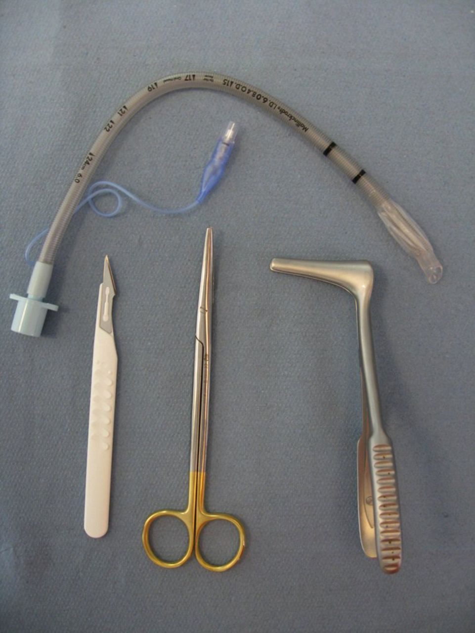
Standard surgical technique (ST) (figure 1),
-
Portex Cricothyroidotomy Kit (PCK) technique (figure 2).

The standard surgical technique set.

The Portex Cricothyroidotomy Kit set.
Each study participant was randomly assigned to one of the cricothyroidotomy technique groups. Furthermore, each study participant performed only one cricothyroidotomy in one cadaver. The standard surgical technique (ST) was performed as follows: (1) Immobilisation of the larynx. (2) Identification of the cricothyroid space followed by a vertical and midline skin incision of 2–3 cm of length using a No. 11 scalpel blade. (3) Blunt preparation of subcutaneous tissue down to the cricothyroid membrane (CTM) with a preparation scissors (eg, Metzenbaum scissors) followed by a transverse incision of the CTM using the same scalpel blade. Withdrawal of the scalpel and use of a Kilian speculum to demonstrate the incision of the CTM. (4) Insertion of a 6.0 mm ID armed cuffed endotracheal tube (Mallincrodt Safety-Flex® low pressure cuff tube; Covidien plc, Dublin, Ireland) with the assistance of a Kilian speculum.
The Portex Cricothyroidotomy Kit (PCK) technique was performed as follows: (1) Immobilisation of the larynx. (2) Identification of the cricothyroid space followed by a vertical and midline skin incision of 2 cm of length only over the CTM. (3) Insertion of the device perpendicularly into the trachea with constant observation of the red indicator in the needle hub. Correct placement into the tracheal lumen is confirmed by disappearance of the red indicator in the needle hub. The device is carefully advanced further until the red indicator reappears again, indicating contact to the posterior tracheal wall. (4) The device is then angled in caudal direction and advanced 1–2 cm into the trachea. The needle is removed and while the dilator is held stationary, the cricothyroidotomy tube is then advanced over the dilator fully into the trachea and the dilator is removed.
In each technique, once the operator had indicated completion of the procedure, correct cricothyroidotomy tube placement was confirmed by anatomical dissection. In accordance with the literature for each technique successful insertion was defined as insertion of the device into the correct anatomic location.5–7 There was no ‘functional check’ by trying to ventilate the patient. During the dissection structures also were inspected for any complications, such as lacerations or penetrations of the tracheal wall, fractures to the trachea, cricoid, or thyroid cartilages. In addition, injury to thyroid vessels were recorded.
After completion of the procedure the following parameters were evaluated: 1. Time to complete procedure (from immobilisation of the larynx to tube insertion). 2. Time to achieve patent airway (from immobilisation of the larynx to tube insertion). 3. Damage to the cricoid or thyroid cartilages, perforation or laceration of the posterior tracheal wall, damage to the oesophagus as well as injury to thyroid vessels.
Statistical analysis of the data was performed using the Fishers exact test to compare success rate and complications. Time to achieve successful airway access was compared using unpaired t-test and reported as mean values (SD). A p value <0.05 was considered statistically significant.
Results
The ST group had a significant higher success rate (100%) when compared to the PCK (67%) group (p=0.04) (see table 1). There was no significant difference in time taken to complete the procedure (p=0.35) as well as to achieve a patent airway (p=0.16) between the two techniques (see table 1). Successful airway access was achieved in a median time of 95 s in the ST and 104 s in the PCK group (p=0.88; see table 1).
Comparison of successful attempts
Complications rates have been higher in the PCK group (67% PCK-group vs 13% ST-group; p=0.04). There was no injury to the thyroid and/or cricoid cartilage (n=0, p=0.04) as well as to the posterior tracheal wall (n=0, p=0.002) in the ST group compared with the PCK group (see table 2). The most frequently encountered complication in the PCK group was injury to the posterior tracheal wall (n=8); out of these eight cases, a laceration of the tracheal wall and a perforation of the posterior tracheal wall as well as the oesophageal wall and penetration into the oesophageal lumen was observed in four cases (see figure 3). In one case a paratracheal tube placement was observed. Therefore, in five cases of the PCK group a patent airway was not achieved (33%). An injury to the thyroid and/or cricoid cartilage was observed in five cases in the PCK group (see table 2). An injury to small vessels of the thyroid glandula was noted in two cases in the ST group (n=2 vs n=0 in the PCK group; p=0.48).
Complications

Severe posterior tracheal wall injury and penetration into oesophageal lumen after tracheal access with the Portex Cricothyroidotomy Kit technique.
Discussion
Emergency cricothyroidotomy is described in the literature as an ‘infrequent’8 or ‘uncommon procedure of which the exact frequency is not known’.9 The incidence of emergency cricothyroidotomy seems to vary considerably depending on a number of factors, like the location of the patient, availability of equipment and skilled assistance as well as individual clinicians' expertise and experience.9–11 Furthermore, emergency cricothyroidotomy is performed often in a ‘cannot intubate—cannot ventilate’ situation in which airway access cannot be obtained by conventional means. Therefore, complication rates from the procedure under these conditions have been reported to be higher (up to 40%).12–16 In order to improve the ease and safety of this emergency procedure, a number of alternatives to the standard surgical technique, such as ‘wire-guided’ and ‘catheter-over-needle’ techniques, have been developed and described in the literature.17 As one of these new devices, the PCK was developed as a ‘catheter-over-needle’ single step insertion technique in conjunction with the UK Military Forces. It is a pre-assembled, compact kit which contains all components for the procedure. In this study we compared this PCK technique with the standard surgical procedure technique in non-formalin-fixated human cadavers, performed by inexperienced clinicians. We studied the three outcome variables success rate, tube insertion time and complication rate, which are crucial for survival of the hypoxic patient. There are several factors influencing these three variables in an emergency situation as well as in a study situation. Out of these factors, familiarity with the device and technique as well as the individual clinicians' experience are of utmost importance.10 Furthermore the type of model (manikin model, porcine cadaver, human cadaver) used for investigation may influence these outcome variables significantly.5 ,10 ,18
In this study the success rate with the indicator-guided PCK device was significantly lower to that with the standard surgical technique (67% vs 100%; p=0.04) caused by paratracheal tube placement (n=1) and oesophageal tube placement (n=4). Success rates in previous studies were variable. Mariappa et al 10 found that only 30% of the attempts with the PCK device on a pig larynx were placed correctly, compared with 55% with standard surgical technique; in this study the operators have been four experienced intensivists with at least 10 years of airway management experience. In contrast to these results, Assmann et al 6 found a success rate of 95% with the PCK device in a study using a standard cricothyroidotomy manikin with an anatomically correct airway. Benkharda et al 19 reported in a study comparing the PCK with a wire-guided technique (Melker Kit) on human cadavers a success rate of the PCK group of 80%; in this study the operators have been two experienced anaesthesiologists. Success rates regarding the standard surgical technique in previous studies using human cadavers vary between 70% and 100%.7 ,18 ,20 ,21 Out of these, the study by Schober et al 18 seems to be most comparable with ours, because of using the same cricothyroidotomy model (human cadavers) and also inexperienced healthcare providers (5th year medical students) performing the procedure; they found a success rate of 94% with the standard surgical technique and 100% with a modified surgical technique (novel scissors technique),18 which is very similar to the success rate in our study (100%).
There was no difference in successful tube insertion time in our study between the PCK-group and the ST-group (median 104 s vs 95 s; NS). We found only one study7 in the literature comparing PCK- and ST-technique. In this study using a porcine airway model no significant difference in the time taken to achieve a patent airway between the PCK and the ST technique was reported and therefore a similar result to our study. In contrast to our findings, previous studies have reported shorter insertion times for the PCK-technique: the median time varies between 33 s6 and 63 s.10 In the only study using human cadavers, a median insertion time of 54 s with the PCK device was reported19; in contrast to our study, in all these studies the operators have been experienced anaesthesiologists or intensivists.6 ,10 ,19 Insertion times for the ST-techniques reported in the literature vary from 32 s22 to 137 s,21 whereas in studies using an artificial or animal airway model, the insertion times have been shorter, than those reported in studies using human cadavers (32–47 s10 ,22 vs 73–137 s7 ,18 ,20 ,21). Out of these studies there is only one, which is comparable with ours regarding airway model (human cadavers), number of attempts (only one per operator) and experience of the operators (inexperienced); with 78 s, Schober et al 18 report of faster insertion times for the ST-technique than we found in our study.
Both the number and the severity of complications and lesions observed were significantly higher with the PCK than with the ST in our study. There was a high incidence of major complications like posterior tracheal wall injury (26.7%) and posterior tracheal wall perforation (26.7%) as well as damage/fracture of thyroid and/or cricoid cartilage (33.3%). Previous studies have reported similar findings: In a study using a porcine airway model, an incidence of 55% of posterior tracheal wall perforation with the PCK-technique was reported.10 In another study using human cadavers, the incidence of ‘major’ complications was 40% including 4/20 (20%) posterior tracheal wall perforations with the PCK-technique.19 There are several reasons for this finding. First, in the PCK device the contact between the needle and the posterior tracheal wall is recommended. Second, the rectilinear and rigid design of the PCK device. Operators in our study reported using a higher insertion force with the PCK device. In accordance with other authors,10 ,19 ,23 we conclude that these factors might have contributed to the high incidence of major complications. We did not experience any of these major complications in the ST-group but two minor complications (injuries to small thyroid vessels). Previous studies have mainly reported similar results. In a large cadaveric study, Schaumann et al 21 did not find any injury to the posterior tracheal wall with the ST-technique. Schober et al 18 in their study did not experience any complication (major and minor) with the ST-technique compared to a complication rate of 36% with a new Scissors-technique and a complication rate of 71% and 64% with a catheter-over-needle and wire-guided-technique. Chan et al 7 also did not experience an injury to the posterior tracheal wall but fractures to the thyroid/cricoid cartilage in 15% of the successful attempts with the ST-technique.
In contrast to manikin as well as animal models, non-fixated, non-frozen fresh human cadavers seem to be the ideal model, allowing standardised research; however, study conditions differ from in vivo conditions. A major difference is that cadavers bleed to a lesser extent so that bleeding complications are likely underestimated.18 This may especially be true for anatomical-surgical techniques and to a lesser extend also to percutaneous puncture techniques because they dilate rather than dissect the tissue. Additionally, laboratory models cannot convey the sense of urgency and difficulty encountered in the clinical situation, where difficult patient anatomy, patient movement, and bleeding are present.24
Conclusions
Our observational bench-test found that the PCK technique was not superior to the ST technique regarding the three outcome variables success rate, insertion time and complications in a human cadaver model performed by inexperienced operators. In contrast, we experienced a significantly lower success rate and a significantly higher rate of major complications with the PCK device compared to the standard surgical technique. In the hand of the inexperienced operator, the standard surgical cricothyroidotomy technique seems to be a safe procedure, which can successfully be performed within an adequate time. The PCK technique cannot be recommended for inexperienced operators.
References
Footnotes
Competing interests None.
Ethics approval Ethics approval was provided by Local ethics committee.
Provenance and peer review Not commissioned; externally peer reviewed.